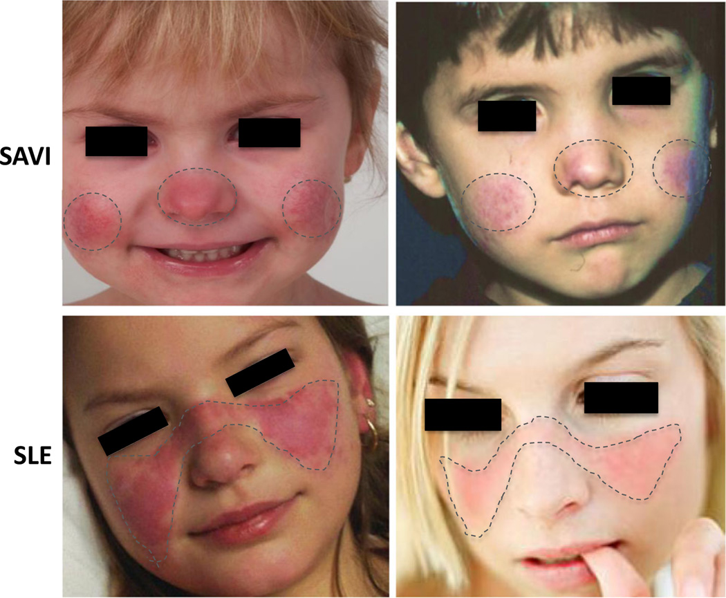
Fig. 1.
Different patterns in facial rash of SAVI and SLE. In SAVI patients (upper panels), the rashes are aggravated by cold exposure and are prominent in cold-sensitive areas including the tip of the nose and the lower part of the cheeks and the ears (not shown). The characteristic malar (facial) rash with a butterfly distribution on pictures from genetically undefined SLE patients (lower panels) show a photosensitive rash that is often induced by exposure to sunlight and associated with immune complex deposition at the dermal-epidermal junction (LE band) on biopsy. References: Lower left photo: © 2016 American College of Rheumatology. Used with permission. Lower right photo: www.mollysfund.org. Used with permission.

