
Figure 2.
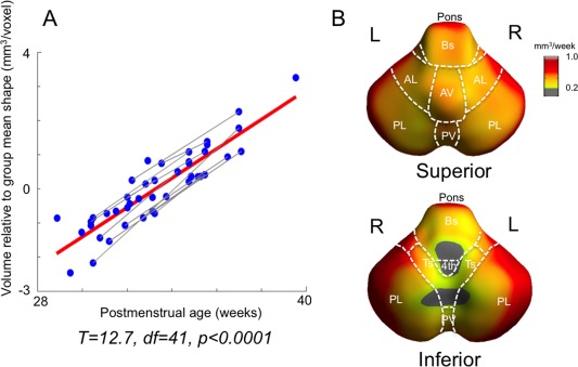
Hindbrain growth in preterm neonates without brain injuries (n = 30). A. Significant mean growth estimated using a mixed‐effect model. Gray lines represent serial scans of individuals, and red line represents overall group correlation. B. Normal growth rates (mm3/wk). Voulme changes are shown within significant areas (corrected P < 0.05). AL/PL: anterior/posterior lobe; AV/PV: anterior/posterior vermis; Ts: Tonsil; Bs: brainstem (medulla); 4th: 4th ventricle; icp: inferior cerebellar peduncle. [Color figure can be viewed in the online issue, which is available at http://wileyonlinelibrary.com.]