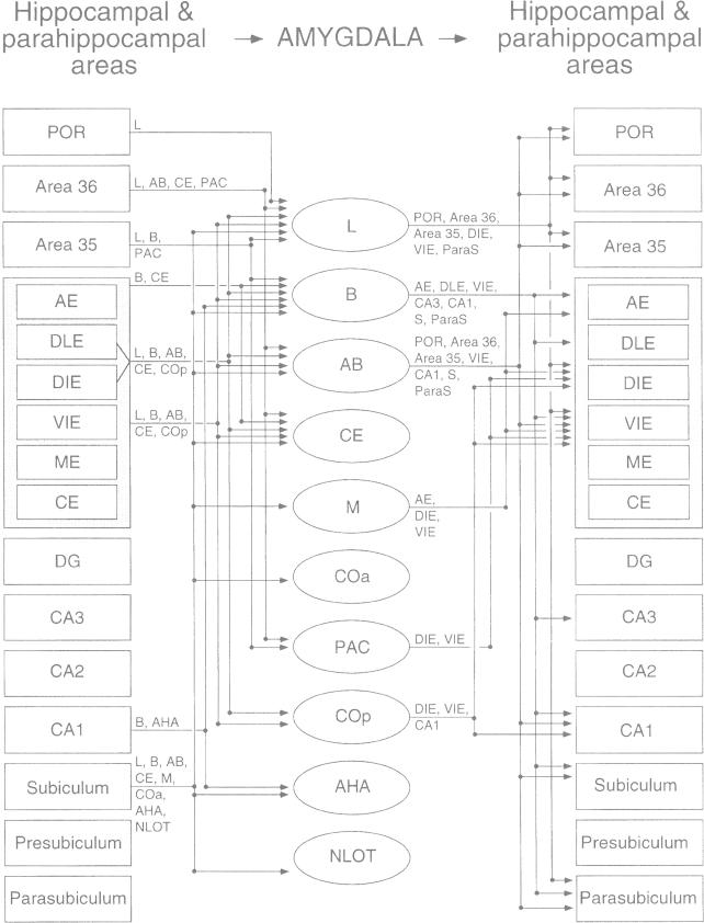
Fig. 2.
Summary of inputs and outputs of the amygdala with the hippocampal region in the rat. The nomenclature used to denote the amygdalar nuclei is that used by Price and coworkers (1987) (see Table 1), and the nomenclature of entorhinal areas (AE, DLE, DIE, VIE, ME, and CE) is that used by Insausti and coworkers (1997) (see text). Only projections that are moderate to heavy in density are illustrated. Abbreviations (with nomenclature of Paxinos and Watson [1997] in parentheses): AB, accessory basal nucleus (posterior basomedial nucleus); AE, amygdalo-entorhinal transitional area (amygdalopiriform transition area); AHA, amygdalohippocampal area; Areas 36 and 35, dorsal and ventral perirhinal areas; B, basal nucleus (basolateral nucleus); CE entorhinal area, caudal entorhinal area; CE amygdalar nucleus, central amygdalar nucleus; COa, anterior cortical nucleus; Cop, posterior cortical nucleus (posteromedial cortical nucleus); DIE, dorsal intermediate entorhinal area (DLEA of Krettek and Price, 1977); DG, dentate gyrus; DLE, dorsal lateral entorhinal area; L, lateral nucleus; M, medial nucleus; ME, medial entorhinal area; NLOT, nucleus of the lateral olfactory tract; PAC, periamygdalar cortex (posterolateral cortical nucleus); POR, postrhinal cortex; VIE, ventral intermediate entorhinal area (VLEA of Krettek and Price, 1977). Reproduced from Pitkänen et al. (2000) with permission.

