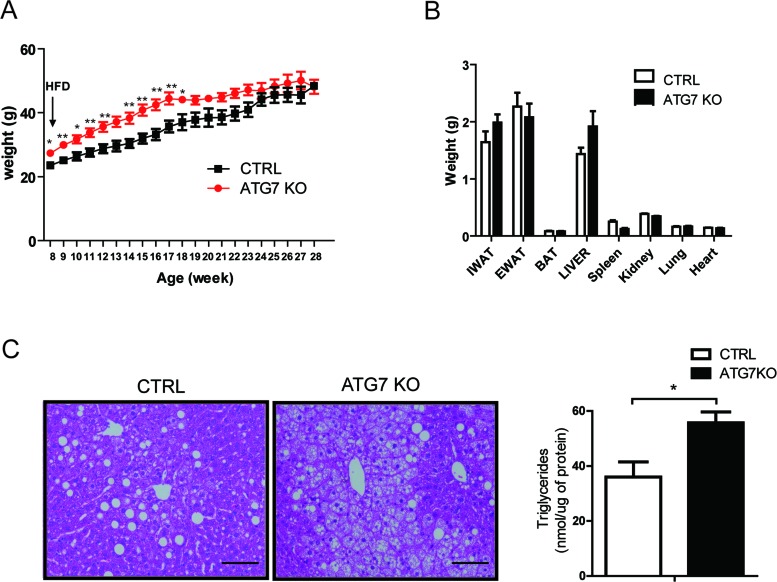
Figure 2. Macrophage-specific Atg7KO mice develop hepatic steatosis under HFD conditions.
A. Atg7fl/fl-LysMCre+/− mice (Atg7KO) (n = 8) and LysMCre+/− mice (control) (n = 6) were fed a 60% HFD, and their growth was monitored by weighing weekly. B. Twenty weeks after administering a 60% HFD, the tissues from Atg7KO (n = 8) and control mice (n = 6) were dissected and weighed. C. Liver sections from Atg7KO and control mice were prepared and stained with hematoxylin and eosin staining. Scale bar, 100 μm (left panel). The triglyceride content of liver tissues from control and Atg7KO mice is shown in the right panel.

