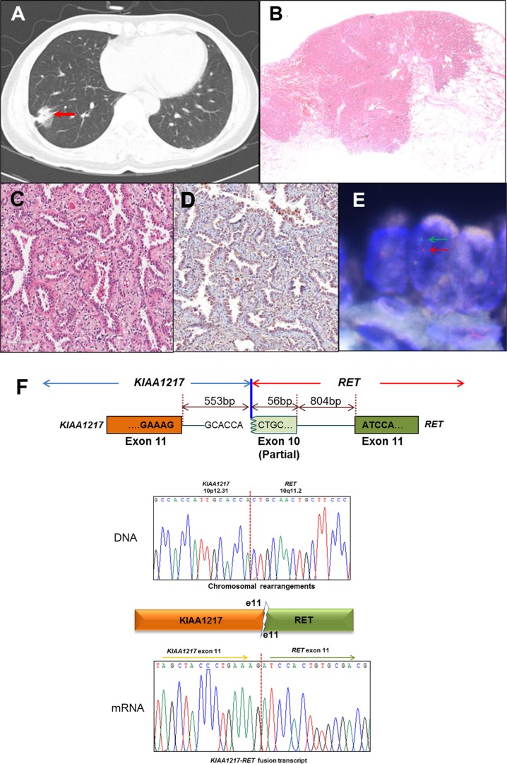
Figure 1. Identification of the KIAA1217-RET fusion gene, (A–E) Clinical and pathological analysis of lung adenocarcinoma harboring RET fusion genes.
(A) A 4-cm solid tumor nodule (red arrow) on the right lower lobe shown by chest computed-tomography scan. (B and C) Histologic features of lung adenocarcinoma harboring RET rearrangement. Adenocarcinoma with a predominant acinar pattern in hematoxylin-and-eosin staining (4 × and 200 ×). (D) Immunohistochemistry of RET shows both membrane and cytoplasm localization (200 ×). (E) Fluorescence in situ hybridization analysis. The split signals (5′-red and 3′-green) were detected in tumor cells. (F) The breakpoints in the KIAA1217-RET fusion gene were identified at the genomic and transcript levels by Sanger sequencing from patient T-#261.

