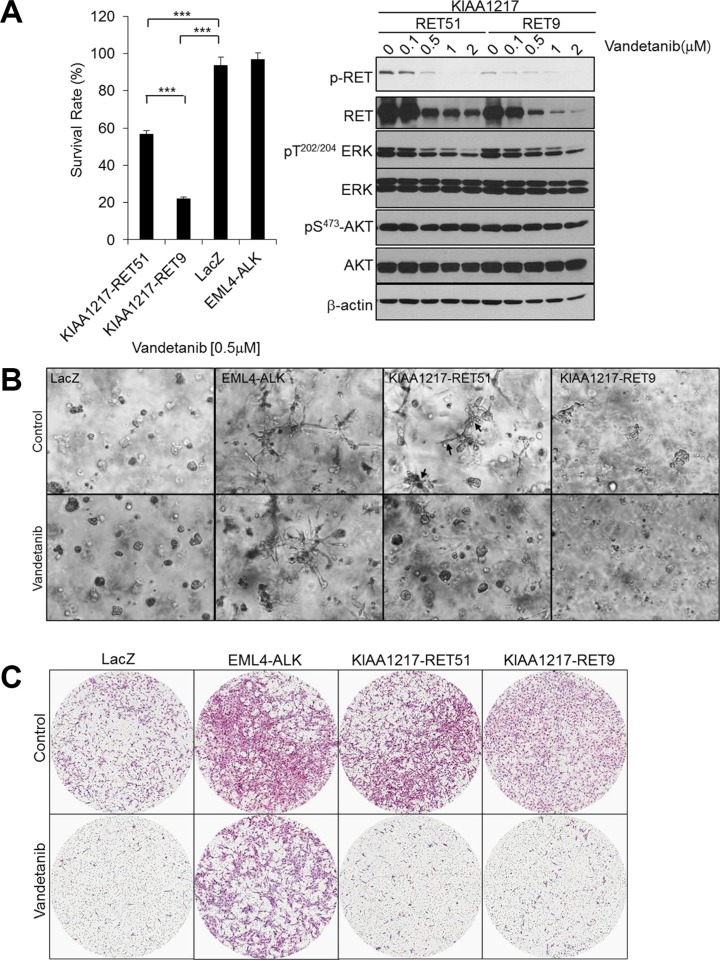
Figure 5. The effects of vandetanib in cells expressing KIAA1217-RET fusion proteins.
(A) Vandetanib reduces proliferation and activation of ERK in cells expressing KIAA1217-RET fusion proteins. (A, left panel) BEAS-2B cells expressing the indicated fusion protein were treated with 0.5 μM vandetanib for 72 h and cell viability was determined. Each bar represents hextuplicate biological replicates ± the standard deviation. ***p < 0.0001. (A, right panel) Cells were treated with the indicated dose of vandetanib for 24 h, followed by cell lysis and detection of the indicated protein by western blot. (B) In vitro colony-forming ability of KIAA1217-RET-positive cells following vandetanib treatment. BEAS-2B cells were embedded in Matrigel and cultured with or without 0.5 μM vandetanib for 7 days. The images were taken by a phase-contrast microscope at 40 × magnification. (C) Invasion abilities of KIAA1217-RET-expressing cells. BEAS-2B cells expressing the indicated protein were loaded onto transwells pre-coated with Matrigel (1 mg/mL), incubated with or without 0.5 μΜ vandetanib for 12 h, and invading cells were fixed and stained with hematoxylin and eosin. The images were obtained using a ScanScope XT slide scanner at 10 × magnification.

