
Figure 8. MIR126 suppresses tumor formation.
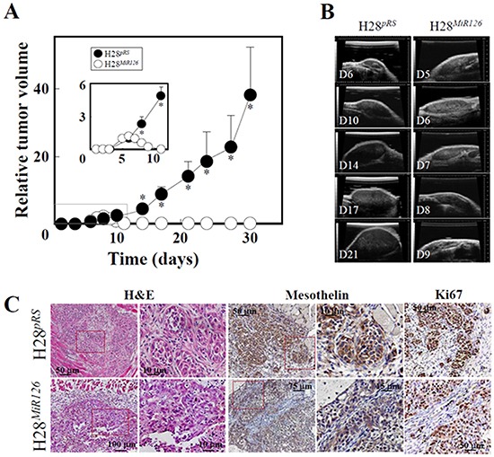
A. Balb-c nu/nu mice were injected subcutaneously with 1×10⁶ H28pRS or H28MiR126 cells and tumor growth was quantified by USI. The inset shows details of tumor size in the initial stage of the experiment. The results are derived from 6 mice in each group, and the symbol ‘*’ indicates significant differences with p<0.05. B. Shown here are representative images of tumors derived from the two H28 sublines acquired on individual days. C. Tumors from day 6 were sectioned and stained with H&E, as well as subjected to immunohistochemistry using anti-mesothelin IgG to identify MM cells in the sections and anti-Ki67 IgG to see the level of proliferation of tumor cells. H&E staining clearly documents lack of ‘structure’ of the H28MiR126 cell-derived tumors, which is obvious in tumors derived from cells transfected with the empty plasmid.