
Figure 3. ZFP91 activates HIF-1α promoter through interacting with transcription factor NF-κB/p65 in colon cancer cells.
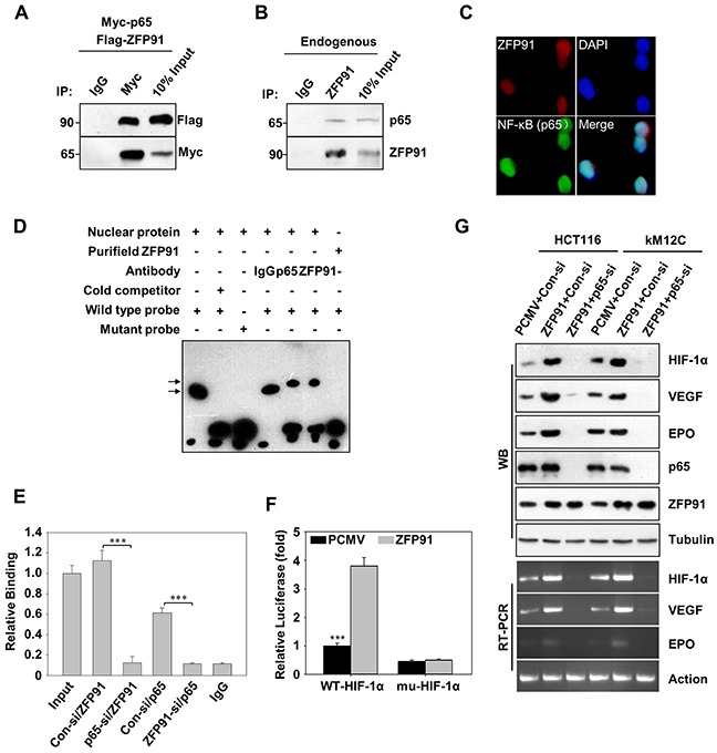
A. HCT116 cells co-transfected with Flag-ZFP91 and Myc-p65 stimulated with hypoxia for final 2 h. Cell lysates were immunoprecipitated with IgG and anti-Myc antibody, and the immune-complexes were analyzed using appropriate antibodies. B. The lysates of HCT116 cells stimulated with hypoxia for final 2 h were incubated with IgG and anti-ZFP91 antibody and the immune-complexes were analyzed using indicated antibodies. C. The nuclear localization of ZFP91 and NF-κB (p65) were detected by immunofluorescence staining in HCT116 cells under hypoxia for final 2 h. The images of ZFP91 (red) and NF-κB (p65) (green) were shown. DAPI staining (blue) was included to visualize the nucleus. D. HCT116 cells stimulated with hypoxia for final 2 h and prepared nuclear extracts for EMSA. The wild type HIF-1α promoter and mutant HIF-1α promoter were used as DNA probes. The shifts (upper arrow) were resulted from the addition of anti-ZFP91 and anti-p65 antibodies. IgG was used as a control antibody. E. HCT116 cells transfected with control siRNA, ZFP91 siRNA or p65 siRNA stimulated with hypoxia for final 2 h. ChIP analysis using antibodies against ZFP91, p65 or non-immune IgG and real-time PCR analysis using specific primers for the HIF-1α promoter and HIF-1α control region was performed. Data represented as mean ± standard deviation of three independent experiments (***p<0.001). F. HCT116 cells co-transfected with PCMV or Flag-ZFP91 together with PGL-WT-HIF-1α or PGL-mu-HIF-1α plasmids stimulated with hypoxia for final 2 h, and then promoter activity of HIF-1α was measured by luciferase reporter gene assay. Data represented as mean ± standard deviation of three independent experiments (***p<0.001). G. HCT116 and KM12C cells pre-treated with PCMV or Flag-ZFP91 together with control siRNA or NF-κB (p65) siRNA stimulated with hypoxia for final 2 h. The cell lysates were analyzed using indicated antibodies. The mRNA expression levels of HIF-1α VEGF and EPO were determined by RT-PCR.