
Figure 5. ZFP91 promotes the tumor growth through HIF-1α in mice.
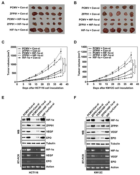
A, B. HCT116 (A) and KM12C (B) cells pre-treated with PCMV, Flag-ZFP91 or HA-HIF-1α together with control siRNA or HIF-1α siRNA were implanted subcutaneously in the left flanks of nude mice. After 40 days, the tumors were dissected out from mice. C, D. The growth curve of tumor. Tumor size was measured every 5 days. Each point showed the mean ± SD (*p<0.05, **p<0.01). E, F. The mean expression levels of HIF-1α, ZFP91, VEGF, and EPO in the tumor tissues from each group mice were determined by western blot and RT-PCR, respectively.