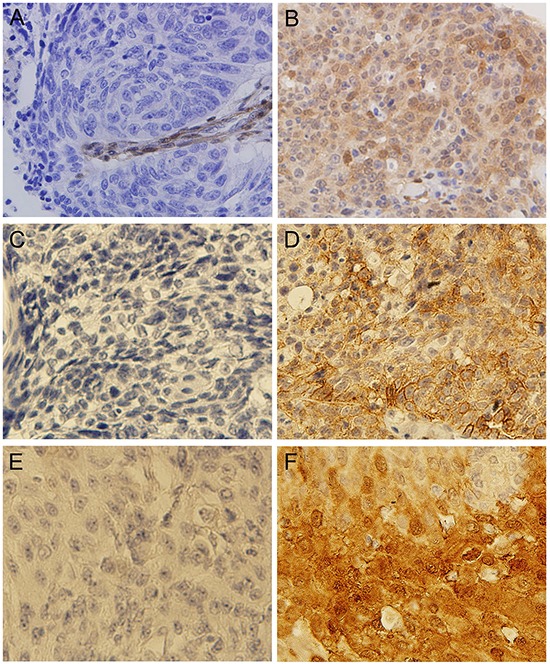
Figure 4. Representative photographs of IHC assay results.

A. Tumor showing lack of staining for PTEN; B. Tumor showing positive staining for PTEN; C. Tumor showing lack of staining for PD-L1; D. Tumor showing positive staining for PD-L1; E. Tumor showing lack of staining for VEGFR2; F. Tumor showing positive staining for VEGFR2.
