
Figure 4. MUC16C promotes cell migration and invasion.
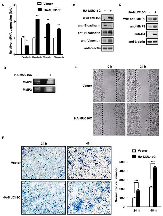
A. MUC16C influences mRNA levels of novel EMT markers like E-cadherin, N-cadherin, Vimentin and Fibronectin. SKOV-3 cells with or without over-expressed MUC16C were harvested at 36 h post-infection and the mRNA levels of above EMT markers were determined by RT-PCR. B. MUC16C influences protein levels of EMT markers. SKOV-3 cells described in (A) were lysed at 36 h post-infection, followed by Western blot with indicated antibodies. MUC16C promotes expression of MMP family members, MMP2 and MMP9, as assessed by Western blot in C. or gelatin zymography assay in D.. SKOV-3 cells for assays in (D) were cultured further in the serum-free medium for another 48 h, and then the culture medium was collected for detecting activated MMP9 and MMP2. E. MUC16C promotes migration of SKOV-3 cells. The SKOV-3 cells described above in (A) were subjected to wound healing assay. Photographs were taken at different time points (0, 24 h). F. MUC16C promotes invasion of SKOV-3 cells. The SKOV-3 cells described above in (A) were subjected to transwell invasion assays. Data are reported as normalized number of cells that invaded through the transwell membrane relative to that of the control (100%), and represent the mean mean±SD of three independent experiments. Significance was calculated by the student's T-test (***P<0.0001).