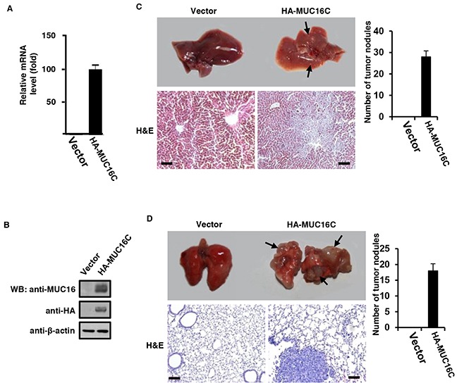
Figure 6. Over-expressed MUC16C enhances metastasis capability of SKOV-3 cells in vivo.

1 × 10⁶ of SKOV-3 cells with or without over-expressed MUC16C were injected intravenously into nude mice (n=5) once a week for consecutive 3 weeks. After another 5 weeks, mice were sacrificed to analyze their main organs and tissues. A. Livers from nude mice described above were homogenated followed by Western blot with antibodies indicated. B. Livers from nude mice described above were homogenated and then the total RNA was extracted for fluorescence quantitative RT-PCR analysis of the RNA level of MUC16/MUC16C. Representative photographs (upper) of livers C. and lungs D. from nude mice described above and the corresponding histological analyses (lower) are shown. Sections were stained with H&E. The black arrows indicate the metastasis stoves. Scale bars 100 μm. Metastatic tumor nodules on the surfaces of livers and lungs were counted and statistical results were presented as histograms in the right (n=5).
