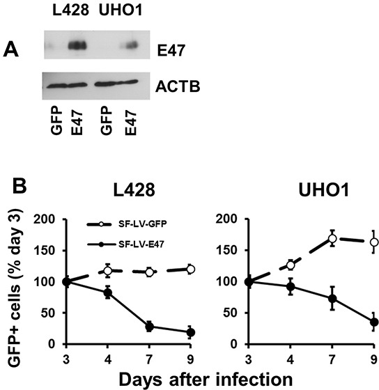
Figure 2. TCF3 negatively regulates proliferation.

E47 was over-expressed in L428 and UHO1 cells using the lentiviral SF-LV-cDNA-EGFP vector. A. E47 protein levels were analyzed by immunoblot. The percentage of GFP+ cells determined by flow cytometry was about 40%. Anti-ACTB antibody was used as a loading control. B. Percentage of GFP+ cells expressing E47 or empty vector (SF-LV) was measured at the indicated time points by flow cytometry. Percentage on the third day of infection (day 3) was defined as 100%. The data shown are representative of three independent experiments.
