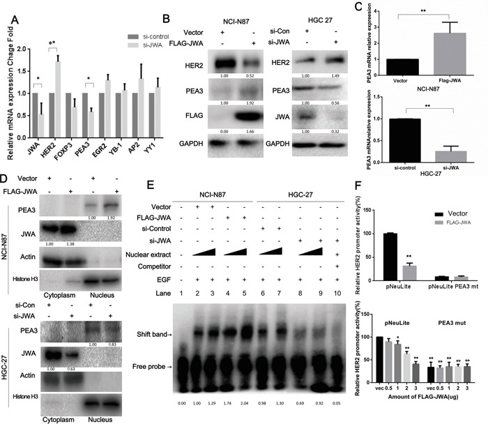
Figure 4. HER2 expression is modulated by JWA through PEA3 upregulation and activation.

A. The mRNA levels of HER2, JWA and a panel of putative transcription factors that regulate HER2 were determined by qPCR after HGC-27 cells were transfected with si-JWA and scramble control RNA. B, C. The mRNA and protein levels of HER2, JWA and PEA3 were identified by qPCR or western blot analyses in NCI-N87 cells transfected with FLAG-JWA or vector as well as in HGC-27 cells transfected with si-JWA or scramble control. * P<0.05; ** P<0.01; Student's t-test. D. PEA3 levels in nuclear and cytoplasmic extracts were confirmed by western blotting in JWA-overexpressing NCI-N87 cells and JWA-silenced HGC-27 cells. Actin and Histone H3 were used as cytoplasmic and nuclear loading controls, respectively. E. Four and six micrograms of nuclear protein were extracted from JWA-knockdown HGC-27 cells and JWA-overexpressing NCI-N87 cells to perform EMSA with a biotinylated oligonucleotide containing the PEA3-binding site and its competitive probe. F. NCI-N87 cells were transiently co-transfected with 2.5 μg (upper panel) or different amounts of FLAG-JWA (lower panel) and 2.5 μg of HER2 luciferase reporter promoter plasmids without (pNeuLite) or with the PEA3-binding site mutation (PEA3mut). The cells were lysed 36 h after transfection, and the luciferase activity was measured. The relative HER2 promoter activity was calculated relative to the activity of the wild-type promoter in vector cells (defined as 100%) after normalization to pRL-CMV. * P<0.05 and ** P<0.01 compared with vector pNeuLite activity.
