
Figure 3. Copper-ionophores generate intracellular ROS and selectively target TRAMP adenocarcinoma cells through a disparity in their antioxidant capacity.
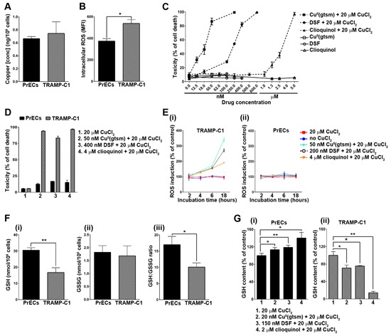
A. TRAMP adenocarcinoma cells (TRAMP-C1) have normal intracellular copper levels. Total intracellular copper was measured in both TRAMP-C1 and mouse primary prostate epithelial cells (PrECs) cultured under basal conditions. Results are shown as copper (ng) per 106 cells. B. TRAMP adenocarcinoma cells (TRAMP-C1) have elevated intracellular ROS levels. Intracellular ROS was measured using the cell permeable fluorogenic probe H2DCF-DA and flow cytometry. Results represent mean fluorescence intensity (MFI) (geometric mean). C. Copper-ionophores potently kill TRAMP adenocarcinoma cells (TRAMP-C1). TRAMP-C1 cells were treated for 18 hours with CuII(gtsm), disulfiram (DSF) or clioquinol alone or in combination with 20 μM CuCl2. Ionophore concentrations are shown and cell viability was determined by the propidium iodide exclusion assay and flow cytometry. D. Copper-ionophores selectively kill TRAMP adenocarcinoma cells while not affecting the viability of mouse primary prostate epithelial cells (PrECs). Both cell lines were treated for 18 hours with CuII(gtsm), disulfiram or clioquinol in combination with 20 μM CuCl2. Ionophore concentrations are shown and cell viability was determined by the propidium iodide exclusion assay and flow cytometry. E. Copper-ionophores generate intracellular ROS in TRAMP adenocarcinoma cells (TRAMP-C1) (i), but not in mouse primary prostate epithelial cells (PrECs) (ii). Both cell lines were treated for 2, 4, 6 or 18 hours with CuII(gtsm), disulfiram or clioquinol in combination with 20 μM CuCl2. Ionophore concentrations are shown and intracellular ROS was measured using the cell permeable fluorogenic probe H2DCF-DA and flow cytometry. Results represent mean fluorescence intensity (MFI) (geometric mean) F. TRAMP adenocarcinoma cells (TRAMP-C1) have markedly reduced antioxidant capacity. Reduced (GSH) (i) and oxidised (GSSG) (ii) glutathione were measured in TRAMP adenocarcinoma cells (TRAMP-C1) and mouse primary prostate epithelial cells (PrECs) by HPLC. (iii) The GSH:GSSG ratio is compared between both cell lines. G. Differential GSH expression in TRAMP adenocarcinoma cells (TRAMP-C1) treated with copper-ionophores. Reduced glutathione (GSH) was measured in mouse primary prostate epithelial cells (PrECs) (i) and TRAMP adenocarcinoma cells (TRAMP-C1) (ii) following treatment for 18 hours with sublethal concentrations of CuII(gtsm) (20 nM), disulfiram (150 nM) or clioquinol (2 μM) (with 20 μM CuCl2). Glutathione (GSH & GSSG) was measured by HPLC. Results represent mean ± STDEV (bar) of triplicate determinations for each measurement. (*p < 0.05; **p < 0.01).