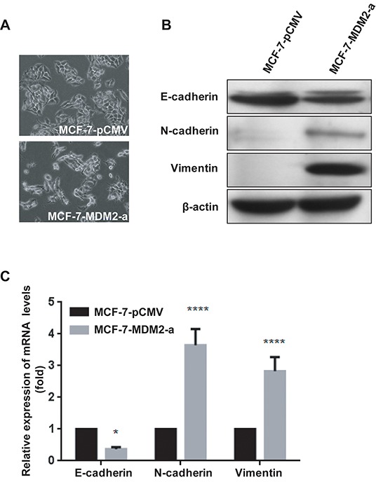
Figure 3. MDM2 overexpression promotes EMT in MCF-7 cells.

Representative phase-contrast images of MCF-7-pCMV and MCF-7-MDM2-a cells showed MDM2-associated morphological changes A. (200×). Expression of epithelial and mesenchymal markers was evaluated by western blotting in MCF-7-pCMV and MCF-7-MDM2-a cells B. β-actin was used as the loading control. Expression of epithelial and mesenchymal markers was analyzed by qRT-PCR in MCF-7-pCMV and MCF-7-MDM2-a cells C. GAPDH served as an internal control. *P<0.05 and ****P<0.0001. The results are from three independent experiments. Error bars indicate the standard deviation.
