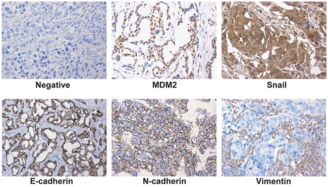
Figure 8. Expression of MDM2, Snail and EMT markers in human breast cancer.

Representative fields of view from the TMA cores show examples of negative staining patterns, positive MDM2, Snail, E-cadherin, N-cadherin and Vimentin (400×). MDM2 exhibited nuclear or cytoplasmic immunoreactivity, Snail exhibited nuclear immunoreactivity; E-cadherin, N-cadherin and Vimentin exhibited membrane or cytoplasmic immunoreactivity.
