
Figure 2. Phosphorylation and activation of PKCδ by Src is important for the scatter of MDCK cells upon HGF stimulation.
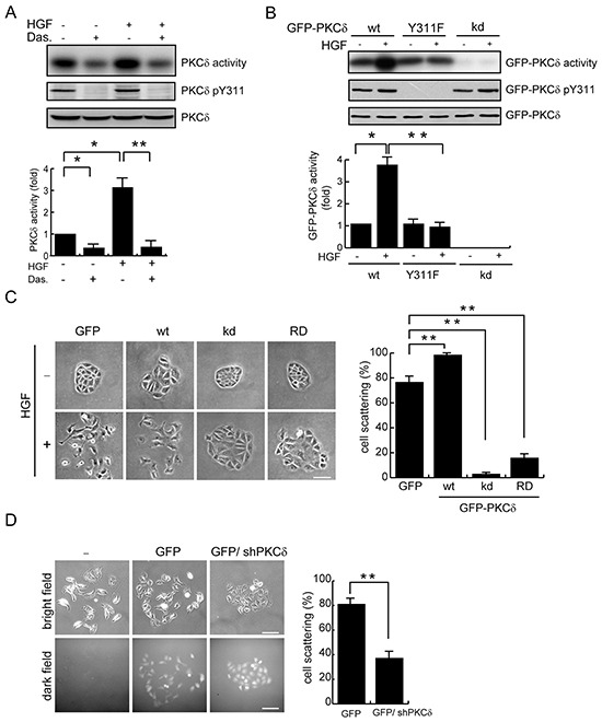
A. MDCK cells were serum-starved for 24 h and were then treated with (+) or without (−) the Src inhibitor dasatinib at 100 nM for 1 h before they were stimulated with HGF (20 ng/ml) for 15 min. Endogenous PKCδ was immunoprecipitated using an anti-PKCδ antibody, and the immunocomplexes were analyzed by immunoblotting with antibodies to PKCδ or PKCδ pY311. To measure the PKCδ activity, the immuoncomplexes were subjected to an in vitro kinase assay in the presence of [γ-32P]ATP and myelin basic protein (MBP) as the substrate. The 32P-incorporated MBP were fractionated by SDS-polyacrylamide gel electrophoresis and visualized by autoradiography. The radioisotope activity was quantified using a phosphoimager system. The data are expressed as fold relative to the level of the control. Values (mean ± SD) are from three experiments. *, P < 0.05; **, P < 0.01. B. MDCK cells stably expressing GFP-PKCδ wild-type (wt), the Y311F mutant, or the kinase-deficient (kd) mutant were serum-starved for 24 h and were then treated with or without HGF (20 ng/ml) for 15 min. GFP-PKCδ was immunoprecipitated by anti-GFP antibody and the immunocomplexes were subjected to an in vitro kinase assay for the PKCδ activity or to immunoblotting with antibodies to GFP and PKCδ pY311. The GFP-PKCδ activity was quantified and expressed as fold relative to the level of the GFP-PKCδ wt. The values (mean ± SD) are from three experiments. *, P < 0.05; **, P < 0.01. C. MDCK cells stably expressing GFP-PKCδ or its mutants were allowed to grow as colonies and were then treated with (+) or without (−) HGF (20 ng/ml) for 12 h to induce cell scattering. The percentage of scattered colonies out of the total counted cell colonies (n ≥ 100) was determined. The values (mean ± SD) are from three experiments. **, P < 0.01. Representative micrographs were taken under a phase-contrast microscope. The scale bar represents 50 μm. D. MDCK cells were transiently transfected with the pSuperior-GFP or the pSuperior-GFP-siPKCδ plasmid that expresses GFP and shRNA specific to canine PKCδ. The cells were allowed to grow as colonies and were then treated with HGF for 12 h. The percentage of scattered colonies out of the total counted cell colonies expressing GFP (n ≥ 100) was determined. The values (mean ± SD) are from three experiments. **, P < 0.01. Representative micrographs of the cell colonies in both bright and dark fields were taken under an epifluorescence microscope. The scale bar represents 100 μm.