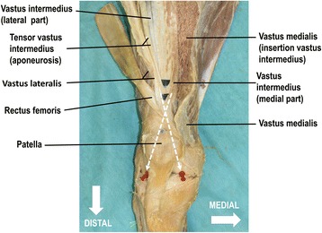
Fig. 2.

Orientation of the multilayered structure of the extensor apparatus of the knee joint. The distal aspect of a right thigh is shown. Red stickers mark the medial and lateral femoral condyles above the knee joint space. The vastus medialis is released from its insertion into the vastus intermedius and rectus femoris (reflected laterally) freeing the view to the complex multi-layered aponeurosis of the vastus intermedius. The lateral part of the vastus intermedius aponeurosis form the deepest layer of the quadriceps tendon. The medial part of the vastus intermedius aponeurosis separates into a superficial and deep medial layer with an orientation towards the lateral femoral condyle (lateral red stick). The fibers of the medial vastus intermedius aponeurosis are located above the lateral part of the vastus intermedius aponeurosis. Generally, lateral fibers are oriented towards the medial femoral condyle (medial red stick). The blue dots mark the fusing points of the intermediate layer of the quadriceps tendon and the superior base of the patella. For better visualization the fusing points are underlined with black paper. The white dotted lines indicate the fiber direction towards the condyles
