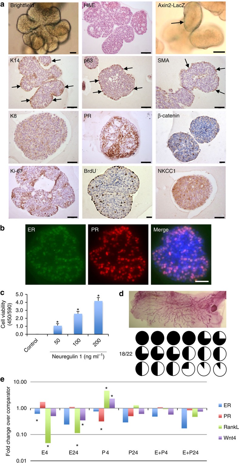
Figure 1. Nrg1 mediates the extended growth of mammary organoids that retain a bilayered organization and response to steroid hormones.
(a) Mammary epithelial cells were embedded in growth-factor reduced matrigel and cultured in basal culture medium supplemented with Noggin (100 ng ml−1) and Nrg1 (100 ng ml−1; n=5). After 30 days in culture, organoids were fixed, embedded in paraffin and sectioned. Sections were stained for hematoxylin and eosin (H&E), basal markers: K14, p63, SMA; luminal markers: K8, PR; cell-proliferation markers: Ki-67 and BrdU; and cell organization markers: β-catenin and NKCC1. Scale bars, 50 μm, except for bright field, β-catenin and BrdU, 25 μm. Mammary organoids established from Axin2-LacZ (Wnt reporter) mammary glands were fixed and stained for β-galactosidase. (b) Detection of ER (green) and PR (red) in mammary organoid section (n=5). Counterstain, DAPI (blue). Scale bar, 50 μm. (c) Mammary cells were exposed to increasing concentrations of Nrg1. After 30 days in culture, the number of viable cells (Wst assay) was measured (n=3, means±s.e.m.). (d) Representative picture of mammary gland successfully filled with a ductal tree. Fifty mammary structures cultured for 30 days were transplanted into mammary fat pads of 3-week-old Rag-1 or FvB mice from which the endogenous epithelium was surgically removed. In all, 18 out of 22 mammary fat pads were re-populated with various degrees (from 5 to 100%). (e) Expression of ER, PR, RankL and Wnt4 following treatment with progesterone (P, 40 ng ml−1), oestrogen (E, 4 ng ml−1) or both hormones. Mammary organoids cultured for 30 days were treated for 4 and 24 h. Gene expression was evaluated by quantitative RT-PCR. Data are expressed as fold change over untreated controls (n=3, means±s.e.m.). *P<0.05, paired Student's t-test.

