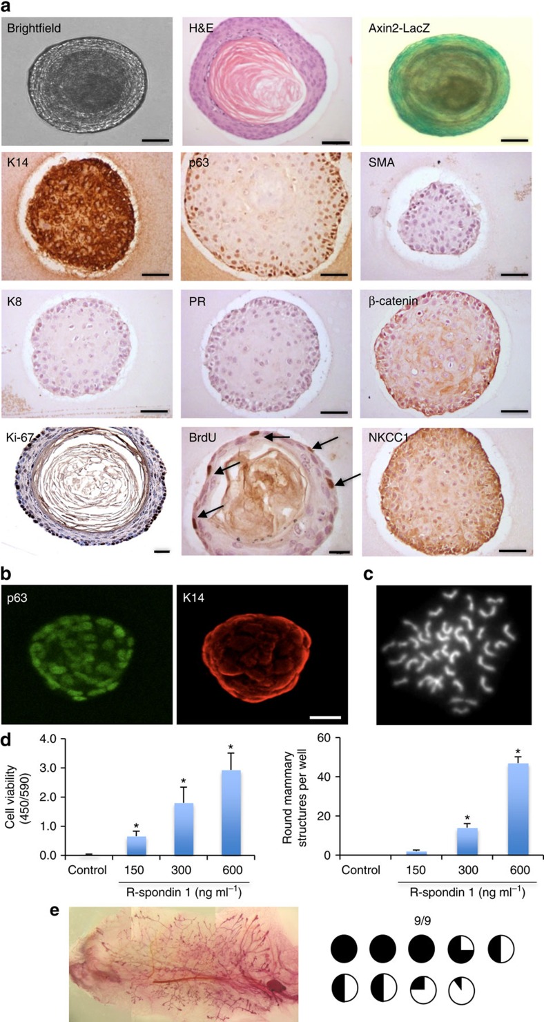
Figure 3. R-spondin 1 promotes the growth of mammary organoids that are enriched in basal cells.
(a) Mammary epithelial cells (n=5) were embedded in growth-factor reduced matrigel. After polymerization of matrigel, basal culture medium supplemented with R-spondin 1 (600 ng ml−1), Noggin (100 ng ml−1) and EGF (50 ng ml−1) was overlaid. After 18 days in culture, organoids were fixed, embedded in paraffin and sectioned. Sections were stained for hematoxylin and eosin (H&E), basal markers: K14, p63, SMA; luminal markers: K8, progesterone receptor (PR); cell-proliferation markers: Ki-67 and BrdU; and cell organization markers: β-catenin and NKCC1. Scale bars, 50 μm, 30 μm (BrdU) and 25 μm (Ki-67). Mammary organoids established from Axin2-LacZ (Wnt reporter) mammary glands were fixed and stained for β-galactosidase. (b) Confocal images of organoids stained for p63 (green) and K14 (red; n=5). Scale bar, 50 μm. (c) Representative picture of metaphase chromosome spread that shows a normal number of chromosomes. (d) Mammary cells were exposed to increasing concentrations of R-spondin 1. After 18 days in culture, cell viability (Wst assay) and the number of structures were evaluated (n=3, means±s.e.m.). (e) Representative picture of mammary gland successfully filled with a ductal tree. Mammary structures cultured for 30 days were transplanted into mammary fat pads of 3-week-old FvB mice from which the endogenous epithelium was surgically removed. In all, 9 out of 9 mammary fat pads were re-populated with various degrees (from 5 to 100%). *P<0.05, paired Student's t-test.

