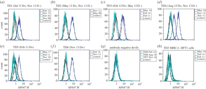
Figure 1.
Flow cytometric analysis of anti-DFT1 antibody responses. (a)–(f) IgG serum antibody results of TD1–6 against MHC-I+ve DFT1 cells compared with negative control. In brackets are the dates each devil was first observed with DFT1 (D+) and when the tumour was no longer present (D−); (g) representative results from three of the 46 devils that had no serum antibody; (h) negative results of TD3 for serum IgG against MHC-I−ve DFT1 cells, representative of TD1–6.

