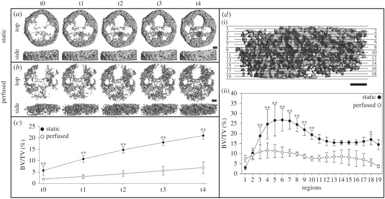
Figure 2.
Three-dimensional images of mineralized tissue formed in the whole scaffold under: (a) static conditions and (b) perfused conditions. (c) Mineralized tissue volume fraction (BV/TV) of the whole scaffold. (d) Schematic illustration of regionalization of the whole scaffold into 10 regions and nine overlapping regions (i) and the corresponding morphometrical profile (ii). Scale bar, 1 mm. *p ≤ 0.05. **p ≤ 0.01.

