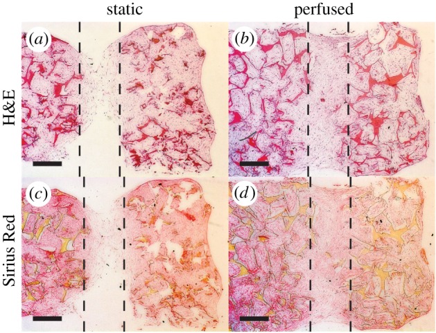
Figure 6.

Vertical scaffold cross-sections stained with haematoxylin and eosin (H&E) (a,b) and Sirius Red (c,d). The dashed lines represent the width of the S channel (1 mm). Cells are located within channels and the pores of the scaffold. Scale bar, 500 µm. (Online version in colour.)
