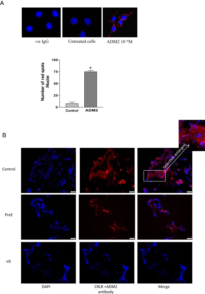
Figure 2.
PLA showing protein-protein interaction between ADM2 and CRLR. A, Increases in the number of red fluorescent dots in ADM2 (10−8 M)-treated cells indicate that ADM2 binds to CRLR in HTR-8/SVneo cells. Bar graph presents the number of spots per nuclei (4′, 6-diamidino-2-phenylindole [DAPI]) that correspond to the number of interactions between CRLR and ADM2. Nuclei staining was done with DAPI. Data are presented as mean ± SEM for three replicate experiments. *, P < .05. B, PLA in the villous explants. Figure shows decreases in the red fluorescence pertaining to ADM2/CRLR association in the villous tissue section from PreE compared with that in the normal placenta (control), thus suggesting PreE-associated decreases in the expression of ligand receptor complex in placenta. Selection in the box is enlarged to show the red fluorescent spots. Absence of antibodies served as negative control (-ve). DAPI was used for nuclear staining (magnification, ×200; n = 3).

