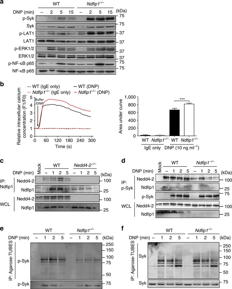
Figure 6. Ndfip1 is an adaptor protein for Nedd4-2 function in mast cells.
(a) Immunoblot analysis of phosphorylated (p-) and total signalling Lyn, Syk, LAT1, ERK1/2 and NF-κB-p65 proteins in whole cell lysates prepared from IgE anti-DNP (SPE-7, 2 μg ml−1) sensitized WT or Ndfip1−/− BMCMCs stimulated with DNP–HSA (20 ng ml−1) for the indicated time points. (b) Representative experiment of DNP induced-Ca2+ influx in WT versus Ndfip1−/− FLMCs (left panel) and quantified analyses of area under the Ca2+ influx curves (right panel). Arrow indicates time of DNP–HSA addition to the cells. Data (mean±s.e.m.) are pooled from the four independent experiments performed, each of which gave similar results. ***P<0.001 for indicated comparison (one-way analysis of variance (ANOVA) with Bonferroni post test). Immunoblot analysis of (c) Nedd4-2 immunoprecipitated (IP) with anti-Ndfip1 from WT and Nedd4-2−/− FLMCs, and (d) Nedd4-2 and Ndfip1 immunoprecipitated with anti-p-Syk from WT and Ndfip1−/− BMCMCs prepared and stimulated with DNP–HSA as in a. above for indicated times. IP: MOCK indicates an IP control performed without antibody. Ubiquitylation of p-Syk (e) and Syk (f) using Agarose-Tandem Ubiquitin Binding Entities in cell extracts from WT and Ndfip1−/− BMCMCs. Data are representative of the three (c–f) independent experiments performed, each of which gave similar results.

