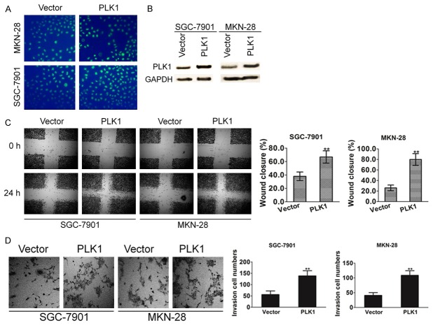
Figure 3.
PLK1 overexpression promotes gastric cancer cell mobility. A. PLK1 was cloned into pIRES2-EGFP vector and then transfected into SGC-7901 and MKN-28 cells. Cells transfected with the empty vector were used as control. The transfection efficiency was evaluated by assayed the green fluorescence protein expression. B. After 24 h post transfections, protein samples were subjected to western blot for measuring PLK1. C. Confluent SGC-7901 and MKN-28 cell monolayers were wounded with 100 µl pipette tip. Cell migration to the wound area was monitored by microscope for 24 h post-wound. The percentage of wound area covered by cells was assessed using the ImageJ program. Bars represent the standard deviation of three independent experiments conducted in triplicate. **P < 0.01 compared with untreated control groups. D. SGC7901 and MKN-28 cells (1 × 10⁵ cells/well) were seeded in the upper chamber, and medium containing 10% FBS was added to the lower chamber. After incubation for 6 h, the cells that invaded the lower membrane of the insert were stained with 0.1% crystal violet and counted by microscopy. The data are expressed as the means of three independent experiments ± standard deviation. **P < 0.01 compared with untreated control groups.

