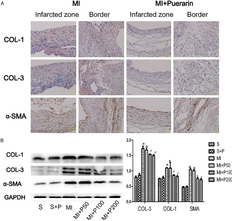
Figure 2.

The deposition of extracellular matrices in the MI myocardium treated with or without puerarin. A. Micrograph of Collagen I, III and α-SMA immunohistochemical staining in MI (left panel) and MI plus high dose puerarin treatment (right panel). B. Representative micrograph of expression of Collagen I, III and α-SMA in westernblot analysis and quantitative results. #P < 0.05 VS MI, *P < 0.05 VS sham group, n=5.
