
FIGURE 1.
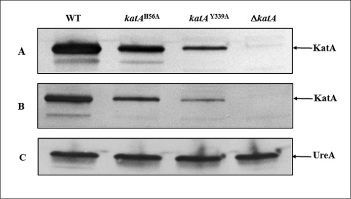
Catalase protein in crude extracts of wild-type and katA mutant strains. Approximately 107 H. pylori whole cells were loaded per lane. Proteins were separated on a SDS-12.5% polyacrylamide gel, along with prestained mass standards, and the proteins were then transferred onto a nitrocellulose membrane and subjected to immunoblotting. Strains are indicated on the top. Proteins detected on each immunoblot are indicated by the arrow on the right. A, anti-KatA immunoblot, strain 43504 (WT) and 43504 katA isogenic mutants. B, anti-KatA immunoblot, strain X47 (WT) and X47 katA isogenic mutants. C, anti-UreA immunoblot, strain 43504 (WT) and 43504 katA isogenic mutants (control to verify equal amounts of cell extracts were loaded in each lane).