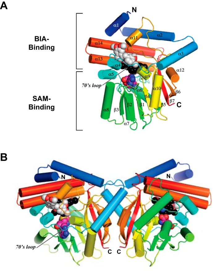
FIGURE 3.

A, overall structure of the PavNMT·SAH·(S)-THP·(R)-THP complex in which the protein is drawn in a ribbon representation, and SAH (carbon atoms colored magenta), (S)-THP (carbon atoms colored black), and (R)-THP (carbon atoms colored light gray) are drawn in a space-filling representation. The polypeptide chain is colored as a gradient from the N terminus (blue) to the C terminus (red). B, dimer structure of the PavNMT·SAH·(S)-THP·(R)-THP complex.
