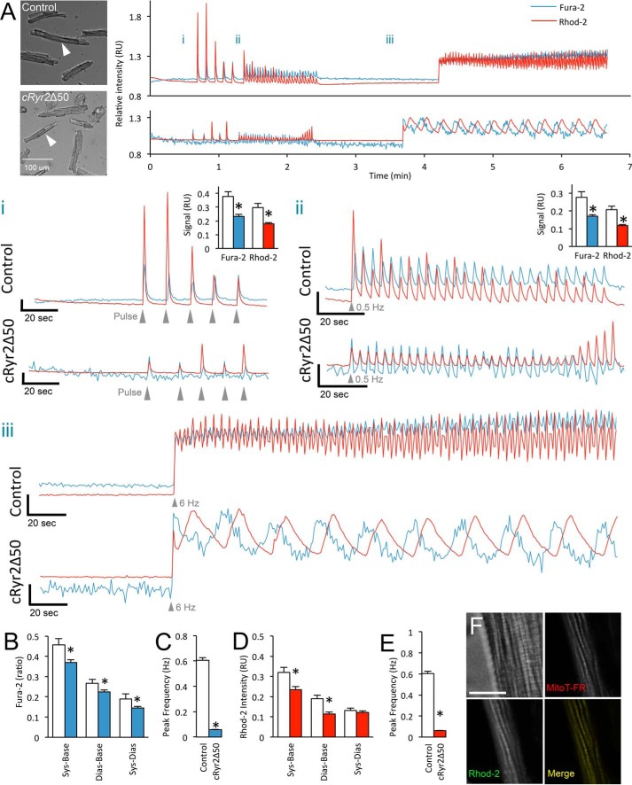
FIGURE 2.
Cytosolic and mitochondrial Ca2+ in cRyr2Δ50 cardiomyocytes. A, representative simultaneous Fura-2 and Rhod-2 fluorescence traces from healthy control and cRyr2Δ50 cardiomyocytes. Bright field images with arrows indicate individual cardiomyocytes from which traces are derived. Panel i, enlargement of pulse stimulation region. Scale axis begins at 0.9 RU. Inset graphs show average peak systolic Fura-2 ratio and Rhod-2 intensity relative to baseline. Panel ii, enlargement of 0.5-Hz stimulation region. Scale axis begins at 0.9 RU. Inset graphs show average peak systolic Fura-2 ratio and Rhod-2 intensity relative to baseline. Panel iii, enlargement of 6-Hz stimulation region. Scale axis begins at 8.5 RU. *, p ≤ 0.05. B, average Fura-2 ratio in control and cRyr2Δ50 cardiomyocytes during 6-Hz stimulation. White bars = control; solid color bars = cRyr2Δ50 throughout (mean ± S.E.). Sys-Base denotes the difference between average peak systolic and baseline measurements, Dias-Base denotes the difference between average minimal diastolic and average baseline measurements, and Sys-Dias denotes the difference between peak systolic and minimum diastolic measurements. *, p ≤ 0.05. C, average frequency of cytosolic Ca2+ transients elicited during 6-Hz stimulation. *, p ≤ 0.05. D, average Rhod-2 intensity in control and cRyr2Δ50 cardiomyocytes during 6-Hz stimulation. *, p ≤ 0.05. E, average frequency of elicited mitochondrial Ca2+ transients observed during 6-Hz stimulation. For all average values: control n = 63 cells, cRyr2Δ50 n = 141 cells, three independent cell preparations from three mice per treatment; *, p ≤ 0.05. F, high resolution microscopy of isolated mouse cardiomyocyte showing colocalization of Rhod-2-AM and MitoTracker Deep Red dye (MitoT-FR). Fluorescent channel images are from a single optical plane from a deconvolved wide field z-stack. Scale bar is 10 μm. All data were plotted as mean ± S.E. Control = Ryr2flox/wildtype + tamoxifen; cRyr2Δ50 = Ryr2flox/wildtype × Mhy6-MerCreMer+ + tamoxifen.

