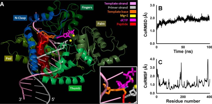
FIGURE 13.
A, MD snapshot at 100 ns of the hPol κ-DNA ternary complex containing the 10-mer peptide cross-linked to 7-deazaguanine. The complex is displayed in schematic mode except for the dCTP and the cross-link-containing template base, which are in stick. The peptide is shown in red surface. The three Watson-Crick hydrogen bonds are shown with dotted lines. The inset, rotated about 90° along the y axis, highlights the Watson-Crick pair distortion. A rotating view is given in the supplemental movie. Cα RMSD (B) and Cα RMSF (C) of the hPol κ over the course of the 100-ns MD simulation for the hPol κ-DNA ternary complex containing the lesion peptide.

