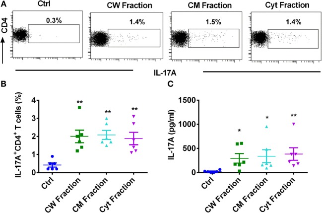
Figure 2.
Different antigen fractions of M. tuberculosis have similar capacity to amplify human monocyte-mediated Th17 response. Monocytes were cocultured with autologous CD4+ T cells either alone or were stimulated with M. tuberculosis-derived cell wall (CW), cell membrane (CM), or cytoplasmic (Cyt) fractions for 5 days. Th17 cells were analyzed by flow cytometry by combination of surface staining for CD4 and intracellular staining for IL-17A. IL-17A in the cell-free supernatants was quantified by ELISA. (A,B) Representative dot plots showing the frequencies of CD4+IL-17A+ T cells and (B) mean ± SEM data from six independent donors. (C) The amount of secretion of IL-17A (mean ± SEM, n = 6). *P < 0.05; **P < 0.01; as determined by one-way ANOVA.

