Figure 3.
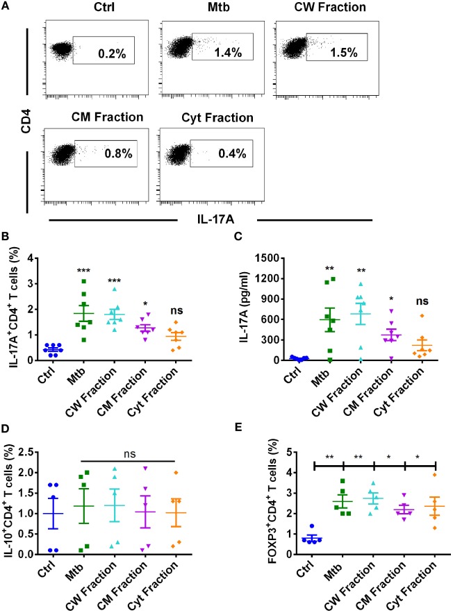
Human dendritic cells differentially promote Th17 response to M. tuberculosis and its antigen fractions. Human monocyte-derived DCs were cocultured with autologous CD4+ T cells at a ratio of 1:10 in X-vivo medium alone or with γ-irradiated M. tuberculosis (Mtb) or M. tuberculosis-derived cell wall (CW), cell membrane (CM), or cytoplasmic (Cyt) fractions for 5 days. Th17 cells were analyzed by flow cytometry by combination of surface staining for CD4 and intracellular staining for IL-17A. IL-17A in the cell-free supernatants was quantified by ELISA. (A,B) Representative dot plots showing the frequencies of CD4+IL-17A+ T cells and (B) mean ± SEM data from seven independent donors. (C) The amount of secretion of IL-17A (mean ± SEM, n = 7). (D,E) Frequencies of IL-10+CD4+ T cells or FoxP3+CD4+ Treg cells in DC–T cell cocultures stimulated with various antigens of M. tuberculosis (mean ± SEM, n = 5). *P < 0.05; **P < 0.01; ***P < 0.001; ns, not significant; as determined by one-way ANOVA.

