
Figure 5.
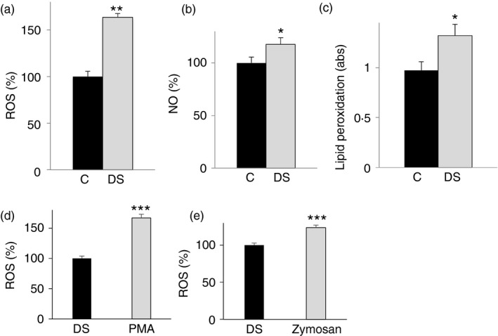
Analysis of oxidative stress in lymphoblasts from children with Down syndrome (DS). Lymphoblastoid cells from six age‐matched control children (black bars) and six children with DS (grey bars) were assayed for reactive oxygen species (ROS) (a), nitric oxide (NO) (b) and lipid peroxidation (c) levels. (d), (e) Lymphoblastoid cells from six children with DS untreated (black bars) or treated (grey bars) with PMA or opsonized zymosan were assayed for superoxide anion radicals by lucigenin assay. In (a), (b), (c), (d) and (e), means ± SD of four replicate independent experiments are shown; where indicated differences between samples and relative controls (*P < 0·05, **P < 0·01) were significant.