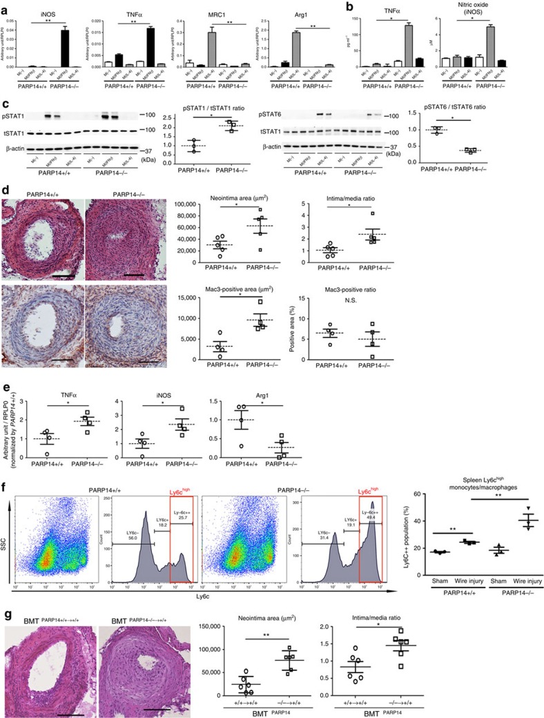
Figure 7. Role of haematopoietic PARP14 in acute arterial lesion formation in mice.
(a–c) Cultured peritoneal macrophages derived from PARP14−/− and PARP14+/+ mice. (a) IFNγ and IL-4 pathway gene expression profiles (n=3). (b) Secretion of inflammatory factors into culture media (n=3). (c) Western blot and corresponding densitometry quantification of phosphorylated STAT1 and STAT6. Each data point is the average of triplicate samples per donor (n=3). (d) Left: representative images of haematoxylin and eosin (H&E; top) and Mac3 (bottom) staining. Scale bars, 100 μm. Right: quantification of lesion formation in mechanically injured femoral arteries of PARP14−/− and PARP+/+ mice. Mac3 staining represents macrophage accumulation (n=4–5). (e) LCM of the neointima followed by gene expression analysis (n=4). (f) Flow cytometry analysis of splenic CD11b+Ly6G− monocytes after induction of mechanically injured femoral arteries of PARP14+/+ and PARP14−/− mice (n=3). (g) Representative H&E staining images and quantification of neointima formation in mechanically injured femoral arteries after bone marrow transplantation (BMT) PARP14+/+→+/+ and PARP14−/−→+/+ mice (n=6). Scale bars, 100 μm. * P<0.05 and **P<0.01, respectively, by Student's t-test. Error bars indicate s.d.

