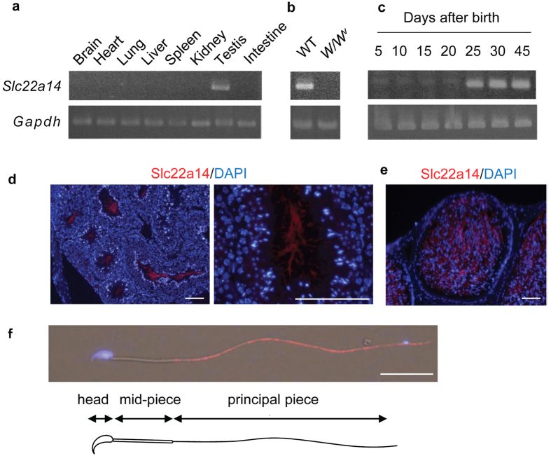
Figure 1. Expression of Slc22a14 in mouse tissues.
(a) Expression analysis of Slc22a14 mRNA in various mouse tissues using RT-PCR. The Slc22a14 signal was detected only in testis. (b) Expression of Slc22a14 mRNA in wild-type (WT) and W/W v mouse testis, which lacks germ cells. (c) Expression of Slc22a14 mRNA during postnatal testicular development. (d) Testicular localisation of Slc22a14. Sections of mouse testis were immunohistochemically stained using anti-Slc22a14 antibody (red). Nuclei were stained with 4′,6-diamidino-2-phenylindole (DAPI, blue). Left panel: lower magnification; right panel: higher magnification. Bar, 50 μm. (e) Immunohistochemical staining of cauda epididymis using anti-Slc22a14 antibody. Bar, 25 μm. (f) Intracellular localisation of Slc22a14 in mouse spermatozoa. Spermatozoa from cauda epididymis was stained as in (d). Slc22a14 is predominantly localised to the principal piece. Bar, 20 μm. The regions of head, mid-piece, and principal piece are shown.

