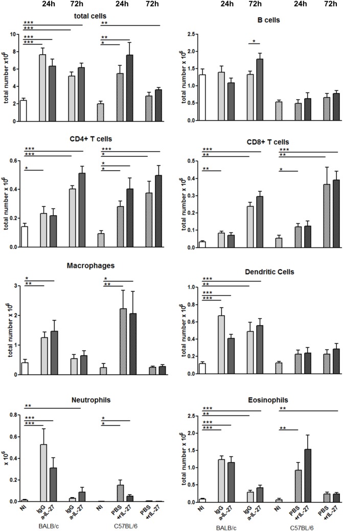
Figure 5.
Cell recruitment to the peritoneal cavity in response to L. infantum infection after IL-27 modulation. BALB/c and C57BL/6 mice were i.p. infected with 1 × 10⁸ promastigotes. Twenty-four hours later, BALB/c mice were treated i.p. with 20 μg of a-IL-27 (clear dark-gray bars) or IgG isotype control (clear light-gray bars), while C57BL/6 received i.p. 1 μg of mouse rIL-27 (patterned dark-gray bars) or the same volume of PBS (patterned light-gray bars). Twenty-four or 72 h after treatment, mice were euthanized and the peritoneal cavity washed. Non-infected (NI) counterparts were always used as controls (white bars, non-patterned for BALB/c and patterned for C57BL/6 mice). Peritoneal cells were then extracellularly stained and acquired by flow cytometry. Bars represent the mean ± SEM of three independent experiments, a minimum of four animals per condition and experiment was analyzed. Unpaired t-test was always used to assess statistical significances (*p ≤ 0.05, **p ≤ 0.01, and ***p ≤ 0.001).

