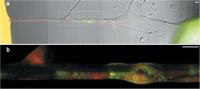
Figure 1. Pseudomonas putida donor and wildtype cells traversing an air gap along a mycelium of Pythium ultimum.
(a) Combined epifluorescence and transmission light image showing mycelium of P. ultimum grown between separate agar pieces inoculated with donor (red) or wildtype (colourless) cells, respectively. Emerging transconjugants (green) are visible along the mycelium. Outlined area is shown in detail in (b). (b) Combined image of red and green fluorescence channels showing the arrangement of donor and transconjugant cells along the mycelial segment. All scale bars represent 10 μm.

