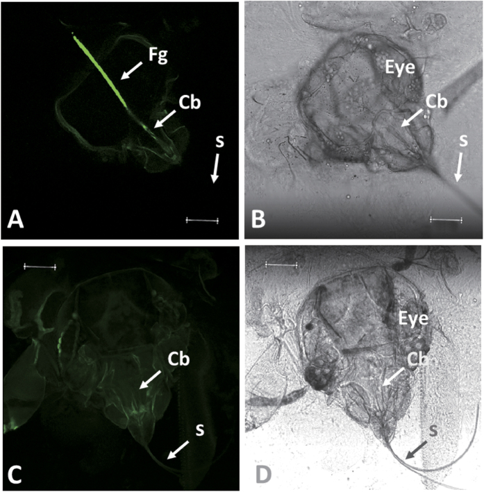
Figure 6. Retention site localization of CCYV virions in the foregut and cibarium of B. tabaci MED under confocal laser scanning microscopy (Leica, TCS SP8).
(A) Confocal view of the head of viruliferous B. tabaci adult, with background transimitted light blocked. (B) Transmitted light view of A. (C) Confocal view of the head of non-viruliferous B. tabaci adult, with background transimitted light blocked. (D) Transmitted light view of C. Under confocal laser scanning microscopy, we observed non-viruliferous (having fed on healthy cucumber plants) or viruliferous whiteflies (having fed on CCYV-infected cucumber plants) after sequential membrane feeding of the following solutions: (i) basal diet, (ii) diet containing anti-CCYV-CP IgG, and (iii) diet containing a goat anti-rabbit IgG conjugated with Alexa Fluor 488. The presence of fluorescent signals showing the retention site within the vector was indicated with the arrow (Fg). The eye, foregut (Fg), cibarium (Cb), and stylets (S) are indicated. (Scale bars, 50 μm).

