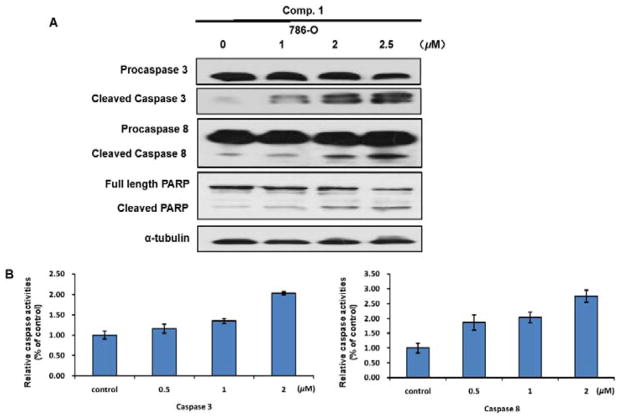
Figure 5.
The apoptosis induced by compound 1 depends on caspase-3 and caspase-8. (A) 786-O Cells were treated with 1, 2, 2.5 μM 1 for 24 h. Cleavage of caspase-3, caspase-8, and PARP was detected by Western blot analysis. α-Tubulin was used as a loading control. A representative blot was shown from three independent experiments. (B) 786-O Cells were treated with 0.5, 1, 2 μM 1 for 24 h. Caspase activation was determined with caspase-3/7 and caspase-8 activity assays.

