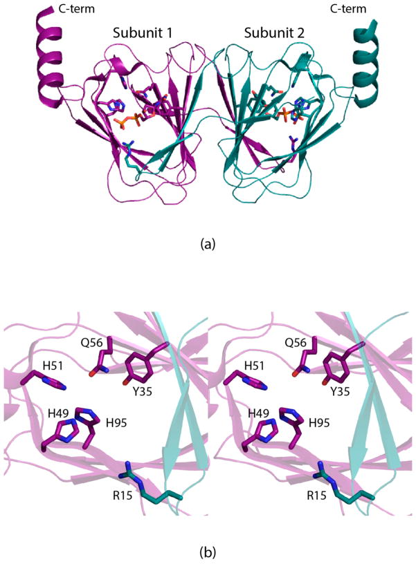
Figure 1.
Structure of FdtA from A. thermoaerophilus (PDB code 2PA7). A ribbon representation of the FdtA dimer is shown in (a) with subunits 1 and 2 displayed in dark violet and dark teal, respectively. The enzyme belongs to the cupin superfamily.18 A close-up view of the active site for subunit 1 is shown in (b). Due to classical domain swapping, Arg 15 is contributed by subunit 2. This figure and Figures 2, 4, 5, 6, and 7 were created with PyMOL.19

