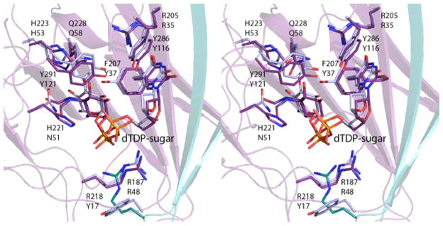
Figure 6.
Comparison of dTDP-sugar binding in the isomerase domain of FdtD and QdtA. The side chains and dTDP-sugar ligands for the FdtD isomerase domain and QdtA are displayed in violet and blue, respectively. The top and bottom residue labels correspond to the FdtD isomerase domain and QdtA. Note that in the wild-type form of QdtA, Asn 51 corresponds to a histidine. In order to trap a dTDP-sugar in the active site, the H51N variant was employed for crystallizations.7

