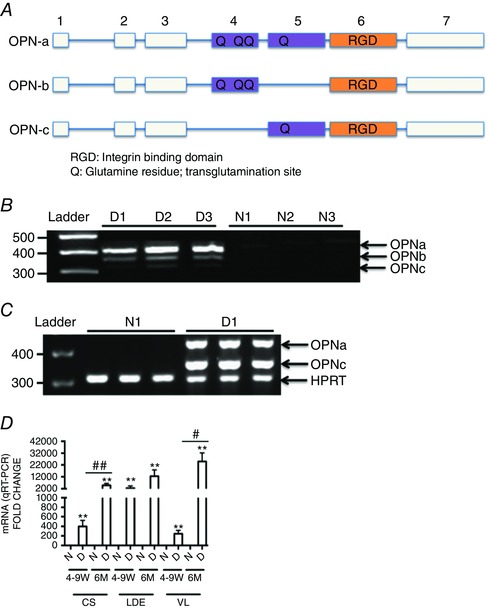
Figure 1. Alternatively spliced osteopontin isoforms are expressed in dystrophic muscle .

A, OPN spliced isoform transcript structure. The blocks in this figure represent exons and the lines represent introns. Here, OPN‐a is full length (top), OPN‐b is missing exon 5 (middle), and OPN‐c is missing exon 4 (bottom). B, osteopontin isoform transcript expression in muscle biopsies of three dystrophin‐deficient Duchenne muscular dystrophy (DMD) patients [D1–D3], and three dystrophin‐sufficient control subjects [N1–N3] as determined by RT‐PCR. Minimal expression is detected in dystrophin‐sufficient muscle, whereas all three isoforms are expressed in DMD muscle (full‐length OPN‐a, OPN‐b lacking exon 5, and OPN‐c lacking exon 4). C, dog dystrophin‐deficient muscle likewise shows no detectable expression in dystrophin‐sufficient littermates (N1), but high expression of OPN‐a and OPN‐c in dystrophin‐deficient golden retriever muscular dystrophy (GRMD) vastus lateralis muscle (D1). D, RT‐PCR analysis of muscle biopsies from three muscles and two age points (4‐9 W, 4–9 weeks old; and 6 M, 6 months old) in dystrophin‐sufficient (N) and ‐deficient (D) dogs shows elevated OPN mRNA in all dystrophic muscles at all age points. An increase in OPN‐a mRNA levels with age is seen in the three muscle groups tested (CS, cranial sartorius; LDE, long digital extensor; and VL, vastus lateralis). Here, OPN‐a mRNA levels were correlated with both age and severity of muscle involvement (CS, mildly affected; and LDE and VL, severely affected). Triplicates are shown per sample. Significant difference between dystrophic and non‐dystrophic littermates: ** P < 0.01. Significant differences with age per muscle group: # P ≤ 0.05 and ## P ≤ 0.01 (GRMD, n = 8; and dystrophin‐sufficient littermates, n = 4).
