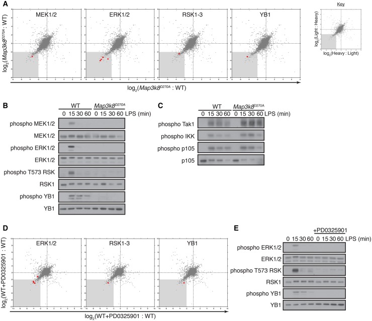
Figure 1. Tpl2D270A mutation inhibits LPS activation of the ERK1/2 and p90RSK.
(A) WT and Map3k8D270A/D270A macrophages, labelled with light and heavy or heavy and light (label reversal) SILAC medium, respectively, were stimulated with LPS for 15 min. Light- and heavy-labelled cell lysates were mixed 1:1 (WT:Map3k8D270A/D270A) based on protein concentration, and phosphosites were quantified by mass spectrometry. Correlation plots show log2 D270A/WT ratios for each phosphosite obtained from the two separate SILAC comparisons. The ratios for the label reversal have been inverted. The grey-shaded area indicates 4-fold down-regulation compared with the control. Phosphosites corresponding to specific components of the ERK1/2 MAPK pathway are indicated with red squares and for clarity are indicated in separate correlation plots. (B and C) WT and Map3k8D270A/D270A macrophages were stimulated with LPS for the indicated times. Total cell lysates were immunoblotted for the indicated antigens. Results are representative of three independent experiments. (D) WT and WT + PD0325901 macrophages, labelled with SILAC medium were stimulated with LPS for 15 min and phosphopeptides analyzed by mass spectrometry as in (A). Correlation plots show log2 WT/WT + PD0325901 ratios for each phosphopeptide obtained from two separate SILAC comparisons, with label reversal ratios inverted. The grey-shaded area indicates 4-fold down-regulation compared with the control. Phosphopeptides corresponding to specific components of the ERK1/2 MAPK pathway are indicated with red squares and for clarity are indicated in separate correlation plots. (E) WT macrophages ± PD0325901 were stimulated with LPS for the indicated times. Total cell lysates were immunoblotted for the indicated antigens. Results are representative of three independent experiments.

