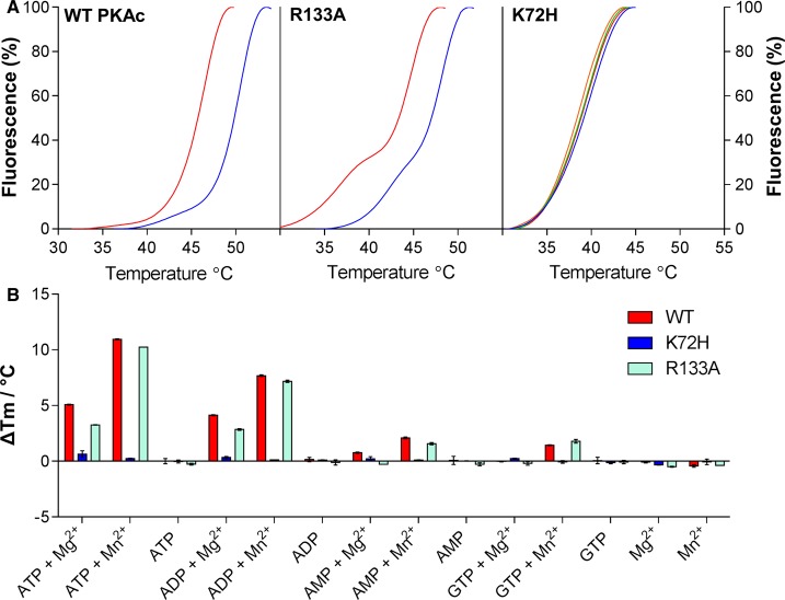
Figure 3. PKA binding of nucleotides is detectable by DSF.
(A) TSA of PKA WT, K72H and R133A (5 μM) in the presence of 10 mM MgCl2 and 1 mM (blue), 2 mM (green) or 4 mM (orange) ATP; buffer control is in red. (B) ΔTm for WT, K72H and R133A PKA upon nucleotide binding, as measured by DSF. Mean ΔTm values ± SD (n = 2) were calculated by subtracting the control Tm value (buffer, no nucleotide) from the measured Tm value.

