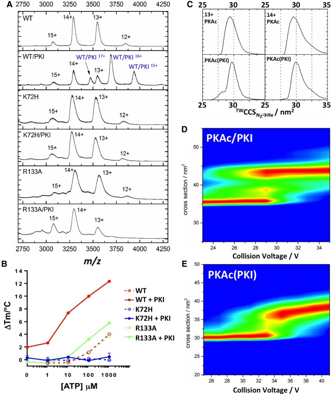
Figure 4. PKI protein binds stably to PKAc WT, but not K72H or R133A protein.
(A) Native ESI mass spectra of PKAc WT, K72H and R133A, in the absence or presence of equimolar PKI. (B) TSA of WT, K72H and R133A PKAc proteins measured in the presence of the indicated concentration of ATP and 10 mM MgCl2 ± 10 μM PKI. Mean ΔTm values ± SD (n = 2) are shown. (C) TWCCSN2→He for [M+13H]13+ and [M+14H]14+ forms of WT PKAc in the absence (top) or presence (bottom) of PKI. CCS distribution of non-PKI-bound form of PKAc is presented [PKA(PKI)]. (D and E) CIU profiles of PKAc upon the addition of PKI: (D) PKI-bound PKA (PKA/PKI) and (E) non-PKI-bound PKAc.

