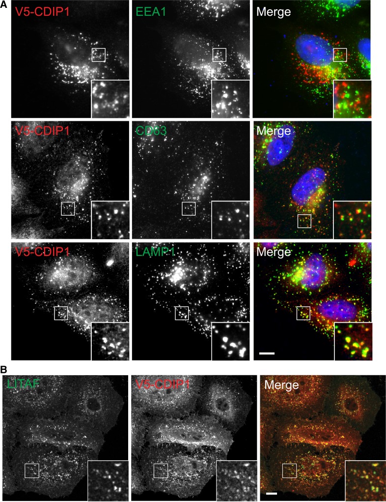
Figure 3. CDIP1 localises to the endocytic pathway.
(A) HeLaM cells expressing V5-CDIP1 were analysed by IF with anti-V5 vs. the indicated endocytic markers. IF is by wide-field microscopy. (B) HeLaM cells expressing V5-CDIP1 were analysed by IF with anti-V5 and anti-LITAF. IF is by confocal microscopy. Scale bars = 10 µm. Insets magnified ×3.

