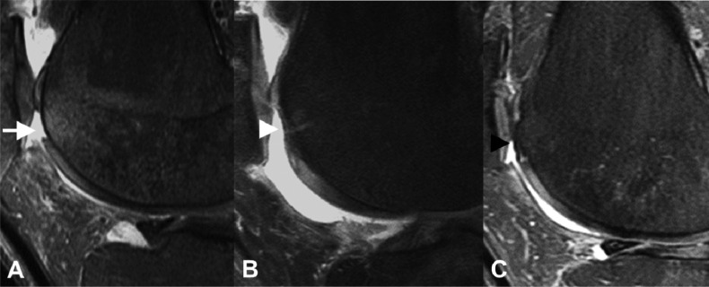
Figure 3.
Trochear microfracture. A) Preoperative sagittal proton density fat-saturated knee MR image shows a full-thickness cartilage defect at the superior aspect of the trochlea with mild subarticular marrow edema (white arrow). B) Post-operative sagittal proton density fat-saturated knee MR image performed 3 months after microfracture demonstrates partial fibrocartilage fill of the microfracture site with minimal residual subarticular marrow edema (white arrowhead). C) Follow-up sagittal proton density fat-saturated knee MR image performed almost 3 years after the microfracture shows near complete fill of the microfracture site with a combination of fibrocartilage and reactive subarticular bone proliferation (black arrowhead). There is complete resolution of the subarticular marrow edema.

