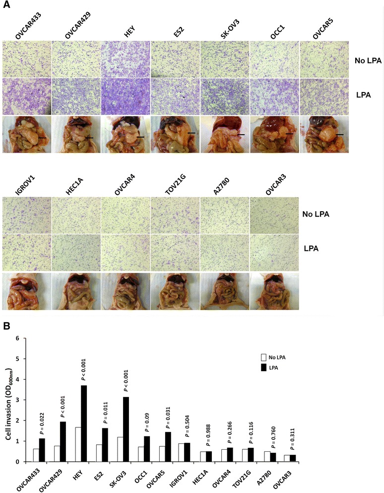
Fig. 1.

Correlation between response to LPA-induced invasion and metastatic colonization potential of ovarian cancer cells. a Invasion of ovarian cancer cells stimulated by LPA. Cell invasion was measured using Matrigel invasion assay with/without 20 μM LPA in the underwells. Peritoneal metastatic colonization assay. The nu/nu mice were intraperitoneally injected with different cell lines (107cells/mice), and autopsied five weeks later. Visible metastatic implants were observed and photographed. b The invaded cells were stained with crystal violet, dissolved in 10 % acetic acid and quantitated with a microplate reader at 600 nm. All samples were performed in triplicate. Data are expressed as the means ± SE
